When there’s a market where patients are less willing to undergo considerable downtime and do not want a significant surgical procedure, the demand for soft-tissue fillers increases. According to data from the American Society for Aesthetic Plastic Surgery (ASAPS), more than 2.1 million hyaluronic acid filler treatments were performed in 2015. That makes hyaluronic acid filler treatments the second most popular nonsurgical cosmetic procedure performed in the United States after neuromodulators; the latter is frequently performed in concert with soft-tissue filler injections.
Physicians have advanced beyond filling rhytides at the forefront of aesthetic medicine developments and are now targeting specific facial sites for deep volumetric augmentation. Evidence for significant volume loss in specific deep fat compartments and facial bones during the aging process is well documented in the medical literature. The aging process occurs in all anatomical layers of the face and rejuvenation should therefore not be limited to dermal signs of aging.
Injection of fillers at strategic target sites can reconstruct youthful anatomy and thereby provide the patient with a natural result. As the relatively new field of aesthetic medicine develops, practitioners are using filler products and techniques with established safety profiles to reduce complications and to increase patient satisfaction. To provide natural-looking results, the physician must have a thorough knowledge of the anatomical changes taking place during the aging process and be able to place fillers in the specific target areas that have lost volume. The practitioner should also be aware of the key anatomical features of each injection site and choose an injection technique that is safe and effective.
The facial arterial system, in particular, represents a danger zone for filler injections, as the intra-arterial injection can potentially lead to widespread necrosis and even blindness. Blindness can occur by injection in an artery in proximity to the orbit and requires rapid specialist intervention. Minimizing the risk of intra-arterial injection of fillers is therefore of paramount importance.
Technological developments include the introduction of the blunt-tipped, or non-traumatic cannula as an alternative to the sharp needle. With a sharp needle, the placement of the needle tip is considered very precise. It is therefore assumed that positioning the tip at the periosteum is relatively easy and will result in precise placement of filler products.
However, the final placement of the filler at this level cannot be guaranteed.
Treatment with non-traumatic cannulae results in significantly fewer bruises and lower pain scores and is gaining in popularity. However, non-traumatic cannulae are more difficult to maneuver to the periosteal level and cannula techniques are therefore wrongly regarded as less precise than those using a needle. Placing the tip of a sharp needle on the periosteum is often thought to be a safe technique to avoid intra-vascular embolization for two main reasons.
First, there are very few arteries running over the periosteum, making it almost an avascular area. Second, it is assumed that a needle with an artery in its trajectory, will completely pierce the artery and exit the other side, avoiding injection of product in the lumen of the artery.
In this cadaver dissection study, our aim was to determine the final position of the injected product using sharp needle vs non-traumatic cannula techniques in a split-face approach. A secondary aim was to study the safety profiles of both injection techniques, related to adjacent arterial danger zones.

INJECTIONS YOUR PATIENTS WILL LOVE! CODE “20OFF” TAKES 20% OFF YOUR FIRST ORDER!
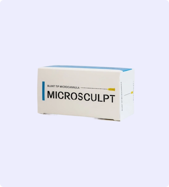
Microcannulas are a tool that every great injector must master. Patients want quick results with no downtime. Our microcannulas are high quality and a fraction of the price of our competitors!
You can create an account here.
How to Minimize Risks of Vascular Occlusion and Blindness During Filler Injections
Dermal fillers improve volume loss or enhance facial features. Their use is increasing at a rate of 10% or more per year worldwide. Adverse events are usually minor and consist of bruising, swelling, asymmetries, and nodularity. More significant complications are fortunately rare and include infection, granuloma, skin necrosis, and blindness. This blog will concentrate on techniques to minimize the risks of having a vascular event.
There are 2 ways a blood vessel can become occluded. If an artery is entered and filler is injected within the lumen (Intraluminal), the filler will travel down the vessel until it gets lodged. At this point, the filler stops the flow of blood to areas that are dependant on this blood supply.
Smaller pieces of the filler can break off and flow into areas far from the initial injection and into the very small arterioles. There are theories that an inflammatory response/cascade exacerbates the injury to the skin and dependent structures. This is Dr. Weiner’s opinion on the etiology of the majority of vascular occlusion cases.
A second way a vessel can occlude is if there is external compression of the vessel by filler. This is plausible in areas of compartmentalization, such as in the nasal tip. If the pressure within the nasal tip exceeds the pressure within an artery, the flow will stop. Unfortunately in this area, vascularity is so poor that peripheral flow doesn’t occur.
External compression is not a major problem in most areas of the face in Dr. Weiner’s opinion. Most vessels can be ligated during surgery and there is no resultant skin necrosis – proving that peripheral flow can make up for an externally compressed vessel.
The worst cases of vascular occlusion result in blindness. This is the result of a filler embolus that travels through an anastomosis between the external and internal carotid systems. The filler backs up into the central retinal artery which feeds the retina. Blood flow is blocked to the retina and blindness ensues.
In most cases, early recognition of a vascular event can be reversed with hyaluronidase if a hyaluronic acid filler was used. Minimal or no sequelae are seen if action is taken within the first 4-6 hours. Unfortunately, even immediate action for blindness related to a filler complication has little or no success.
There have been about 100 reported cases of blindness from fillers, with most of the cases coming out of Asia. This is certainly underreported though. The areas of most risks for blindness are injections in: glabella, nose, periocular, and NLF. Fat is the most common filler causing blindness, but all fillers have been implicated. Any area of the face is at risk for vascular occlusion/necrosis.
Ninety-eight cases of vision changes from filler were identified. The sites that were high risk for complications were the glabella (38.8%), nasal region (25.5%), nasolabial fold (13.3%), and forehead (12.2%). Autologous fat (47.9%) was the most common filler type to cause this complication, followed by hyaluronic acid (23.5%). The most common symptoms were immediate vision loss and pain. Most cases of vision loss did not recover. Central nervous system complications were seen in 23.5% of the cases. No treatments were found to be consistently successful in treating blindness.
Techniques for optimizing safety during dermal filler administration:
- Know the major vascular structures and their landmarks
- Avoid areas you (the injector) are not comfortable with. Particularly the high risk areas: glabella, nose, periocular
- Consider using only reversible fillers if there is any concern regarding vascular occlusion or experience
- Use cannulas whenever feasible, preferably 25/23g or larger
- Avoid boluses, small linear threads are safer
- Constantly move tip of cannula/needle. If more filler is needed in a particular area, revisit the area with another pass.
- A NEGATIVE ASPIRATION DOESN’T EQUATE TO BEING EXTRAVASCULAR AND CAN GIVE A FALSE SENSE OF SAFETY
- Injection onto periosteum is safest but does not guarantee a vascular free injection
- Pressure on the supratrochlear vessels during glabellar or nasal injections might limit reflux of filler into the orbital vessels
- Retrograde injections are safer than anterograde injections
- Dermal injections should be relatively safe
- Avoid deep injections in the lips. Stay superficial to the muscles
- An injection that is perpendicular to a vessel is purported to be safer than one which is parallel because the time within the vessel should be less if it is entered
- Have on hand 6-8 vials of Hylenex
- Any unusual bruising, pain or visual change needs immediate evaluation
The bottom line is that complications can occur with dermal fillers, even during a routine procedure. Use of larger gauge microcannulas greater than 25 gauge may minimize the risk of vascular complications but does not make it zero. Many measures can be taken to minimize risks. Choosing an experienced injector will result in safer and better outcomes.
Sources
- Beleznay, Katie MD, FRCPC, FAAD*; Carruthers, Jean D. A. MD, FRCSC, FRC (OPHTH), FASOPRS†; Humphrey, Shannon MD, FRCPC, FAAD*; Jones, Derek MD‡,§ Avoiding and Treating Blindness From Fillers, Dermatologic Surgery: October 2015 – Volume 41 – Issue 10 – p 1097-1117 doi: 10.1097/DSS.0000000000000486
- Beleznay K, Carruthers JD, Humphrey S, Jones D (2015) Avoiding and treating blindness from fillers: a review of the world literature. Dermatol Surg 41:1097–1117
- Youn SH, Seo KK (2016) Filler rhinoplasty evaluated by anthropometric analysis. Dermatol Surg 42:1071–1081
-
 23 gauge 50 mm (2 inch) Microcannulas
 -
 22 Gauge 100 mm (4 inch) Microcannulas.
 -
 27 Gauge 38 mm (1.5 inch) Microcannulas
 -
 25 Gauge 38 mm (1.5 inch) Microcannulas
 -
 30 Gauge 25 mm (1 inch) Microcannulas
 -
 18 Gauge 100 mm (4 inch) Microcannulas
 -
 25 Gauge 50 mm (2 inch) Microcannulas
 -
 21 Gauge 50 mm (2 inch) Microcannulas
 -
 21 Gauge 70 mm (2.75 inch) Microcannulas


