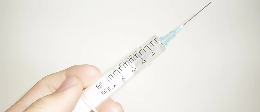
 06/20/2020In a recent study conducted by The People's Republic of China, multiple doctors tested various types of microcannulas to measure their…
 03/10/2020Why Do Physicians love Microcannulas? Simply put? The clinical benefits. Benefits from blunt microcannulas include elimination or near elimination of…
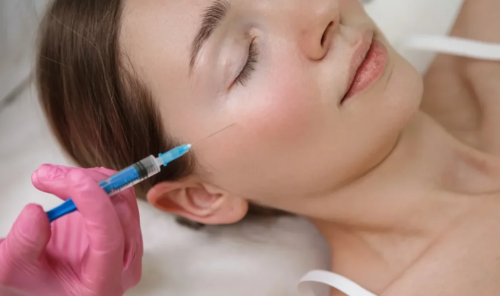
 03/10/2020For decades dermal fillers have been delivered to the face through a hypodermic needle. Nevertheless, aesthetic cannulas have become more…